Anatomy of Facial Expression
The human face is a complex and expressive organ, capable of conveying a wide range of emotions. This ability is made possible by a intricate network of muscles that work together to create subtle and nuanced movements. Understanding the anatomy of facial expression is crucial for artists, animators, and medical professionals who seek to accurately portray and interpret human emotions.
Introduction
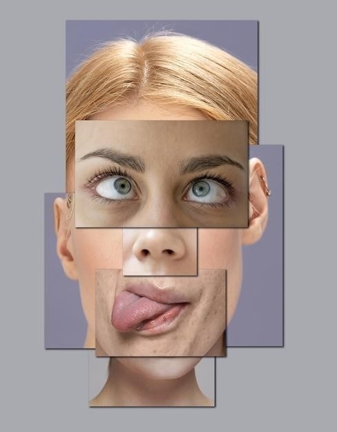
Facial expressions are a fundamental aspect of human communication, playing a vital role in conveying emotions, intentions, and social cues. The ability to recognize and interpret these expressions is essential for effective social interaction. The anatomy of facial expression encompasses the intricate network of muscles, nerves, and structures that contribute to the dynamic movements of the face. This anatomical framework, when understood, allows for a deeper appreciation of the complexity and nuances of facial expression.
This understanding is not only crucial for artists and animators seeking to portray realistic and expressive characters but also for medical professionals who diagnose and treat facial disorders. The study of facial expression anatomy offers valuable insights into the interplay between physical structures and emotional states, providing a foundation for understanding the language of the human face.
Facial Muscles⁚ The Building Blocks of Expression
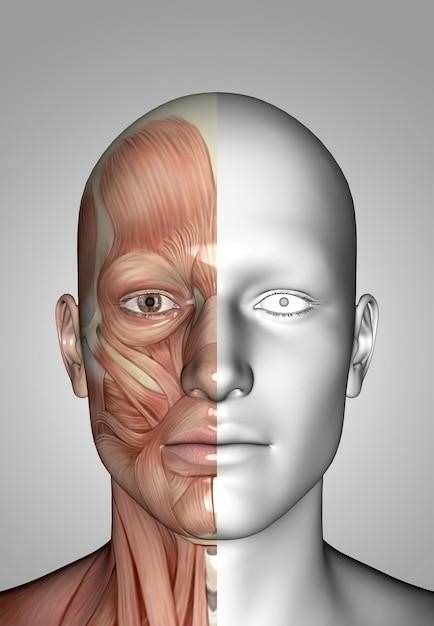
The muscles of facial expression, also known as mimetic muscles, are the primary architects of our facial movements. These muscles are unique in that they attach to the skin rather than bones, allowing for a wide range of subtle and dynamic movements. They are responsible for creating the expressions that convey our emotions, from a simple smile to a complex grimace. The intricate interplay of these muscles, each with its specific function, gives the face its remarkable ability to communicate a vast spectrum of feelings.
These muscles are strategically positioned throughout the face, and their coordinated contractions and relaxations generate the characteristic movements associated with different expressions. The ability to control these muscles allows us to communicate nonverbally, adding depth and nuance to our interactions. Understanding the anatomy of these muscles is key to appreciating the intricate mechanisms behind facial expressions and their role in human communication.
Types of Facial Muscles
The muscles of the face can be broadly categorized into two primary groups⁚ mimetic muscles and mastication muscles. Mimetic muscles, as their name suggests, are primarily responsible for facial expressions and are the key players in conveying emotions. They are delicate and intricate, attached to the skin, and their contractions create the subtle movements that shape our facial expressions.
In contrast, mastication muscles, also known as chewing muscles, are larger and more powerful. They are responsible for the complex movements of the jaw, essential for chewing and speaking. These muscles attach to the bones of the skull and jaw, providing the strength and stability necessary for these essential functions. While not directly involved in facial expressions, they play a crucial role in the overall function and appearance of the face.
Mimetic Muscles
Mimetic muscles are the primary actors in the theatre of human emotion; They are the delicate, intricate muscles that attach to the skin, allowing for the subtle movements that shape our facial expressions. These muscles are responsible for the myriad of expressions we display, from the subtle twitch of a smile to the full-blown contortions of anger or fear.
Each mimetic muscle has a specific role to play in creating a particular expression. For example, the orbicularis oculi muscle, which surrounds the eye, is responsible for closing the eyelid and creating the characteristic squint of suspicion or concentration. The zygomaticus major muscle, which runs from the cheekbone to the corner of the mouth, is responsible for pulling the lips upwards, creating the smile that signals happiness and amusement.
Mastication Muscles
While mimetic muscles are the stars of the emotional show, mastication muscles are the unsung heroes of everyday function. These muscles are the powerhouse of the jaw, responsible for the complex actions of chewing and biting. They are larger and stronger than mimetic muscles, designed for the heavy lifting of breaking down food.
The primary mastication muscles are the masseter, temporalis, medial pterygoid, and lateral pterygoid. These muscles work together to elevate, depress, protrude, and retract the mandible, allowing for the precise movements needed to grind and crush food. The masseter, a powerful muscle located on the side of the face, is responsible for closing the jaw, while the temporalis, located on the temporal bone, assists in this action and also helps to retract the jaw. The medial pterygoid also helps to close the jaw, while the lateral pterygoid is responsible for opening the jaw and moving it from side to side.
Innervation of Facial Muscles
The intricate dance of facial muscles is orchestrated by a complex network of nerves, ensuring precise control over every twitch and expression. The facial muscles are innervated by the facial nerve (VII), a cranial nerve that emerges from the brainstem and travels through the skull to reach its target muscles.
The facial nerve carries both motor and sensory fibers, allowing it to control muscle movements and relay sensory information from the face. Its motor fibers innervate all the muscles of facial expression, enabling us to smile, frown, raise our eyebrows, and perform a myriad of other facial gestures. The sensory fibers of the facial nerve are responsible for conveying taste sensations from the anterior two-thirds of the tongue and providing sensory feedback from the skin of the face and ear.
The intricate innervation of the facial muscles allows for a remarkable degree of control over our expressions. This delicate interplay between nerves and muscles is what gives our faces their dynamic and expressive nature.
The Facial Nerve (VII)
The facial nerve (VII), also known as the seventh cranial nerve, is a crucial player in the symphony of facial expressions. It acts as the conductor, transmitting motor commands from the brain to the muscles of the face, allowing us to convey a wide range of emotions. This complex nerve emerges from the brainstem, a region at the base of the brain, and embarks on a journey through the skull, passing through a narrow opening called the stylomastoid foramen.
Once it exits the skull, the facial nerve branches out, sending its motor fibers to innervate all the muscles of facial expression. This intricate network of nerve branches ensures precise control over every subtle movement of the face, from a gentle smile to a wide-eyed expression of surprise. The facial nerve also carries sensory fibers that relay taste sensations from the anterior two-thirds of the tongue and provide sensory feedback from the skin of the face and ear.
The facial nerve’s crucial role in facial expression is evident in conditions like Bell’s palsy, where damage to the nerve leads to facial paralysis, significantly impacting a person’s ability to communicate and express emotions. Understanding the anatomy and function of the facial nerve is essential for medical professionals to diagnose and treat disorders that affect facial expression.
Clinical Significance
The anatomy of facial expression holds significant clinical importance, as it provides insights into various medical conditions and their impact on human health. Understanding the intricate network of muscles and nerves responsible for facial movements enables medical professionals to diagnose and treat a range of disorders affecting facial expression. This knowledge is crucial for neurologists, plastic surgeons, and other specialists who work with patients experiencing facial paralysis, muscle weakness, or other conditions that affect their ability to communicate emotions and interact with the world.
For instance, understanding the anatomy of the facial nerve is essential for diagnosing and managing Bell’s palsy, a condition characterized by sudden facial paralysis due to inflammation of the facial nerve. Additionally, knowledge of the facial muscles and their innervation is critical for planning and performing surgical procedures such as facial reconstruction, nerve grafts, and muscle transfers, aimed at restoring facial function and improving quality of life for patients with facial paralysis or other facial deformities.
Furthermore, the study of facial expression provides valuable insights into neurological disorders, such as Parkinson’s disease and stroke, which often manifest as changes in facial movements and expressions. By understanding the anatomy and function of the facial muscles and nerves, medical professionals can better assess the severity and progression of these conditions and develop appropriate treatment strategies.
Facial Paralysis
Facial paralysis, also known as facial palsy, is a condition that affects the ability to move the muscles on one or both sides of the face. This condition can be caused by a variety of factors, including nerve damage, stroke, tumors, or infections. The most common cause of facial paralysis is Bell’s palsy, a temporary condition that affects the facial nerve.
Facial paralysis can significantly impact a person’s ability to communicate, eat, and even breathe. It can also lead to social isolation and psychological distress. The severity of facial paralysis can vary widely, from mild weakness to complete paralysis. Treatment for facial paralysis depends on the underlying cause and may include medications, physical therapy, or surgery.
Physical therapy exercises can help strengthen the affected muscles and improve facial movement. In some cases, surgery may be necessary to repair damaged nerves or to transfer muscles to restore facial function. The prognosis for facial paralysis depends on the underlying cause and the severity of the condition. With timely and appropriate treatment, many people with facial paralysis can experience significant improvement in their symptoms.
Applications in Art and Animation
The study of facial expression anatomy is invaluable for artists and animators who strive to create realistic and expressive characters. By understanding the interplay of muscles and their corresponding movements, artists can accurately depict a wide range of emotions, from subtle nuances to intense expressions.
Knowledge of facial muscle anatomy allows artists to sculpt, draw, or paint faces with greater precision and realism. This understanding enables them to depict wrinkles, creases, and subtle muscle contractions that contribute to the overall expression. Animators, on the other hand, utilize this knowledge to create believable and engaging character animations. They can program realistic facial movements, expressions, and reactions, enhancing the emotional impact of their creations.
The study of facial expression anatomy serves as a foundation for artists and animators, allowing them to create characters that resonate with viewers on an emotional level. It empowers them to bring life and depth to their creations, making them more captivating and relatable.
The intricate network of facial muscles, their innervation, and their coordinated movements form the foundation of human facial expression. Understanding this intricate system is essential for various disciplines, including art, animation, and medicine. Artists and animators leverage this knowledge to create realistic and expressive characters, enhancing their creations’ emotional impact. Medical professionals utilize this understanding to diagnose and treat facial paralysis, improving patients’ quality of life.
The study of facial expression anatomy continues to evolve, with advancements in technology and research providing deeper insights into the complex mechanisms of facial movement. This knowledge empowers us to better understand and appreciate the nuances of human expression, fostering greater empathy and communication within our society.